Pairing OCR and the ECAR metabolic assays for a comprehensive characterization of metabolic activity in Ncardia’s human iPSC-derived cardiomyocytes.
The combination of these two metabolic assays with physiologically relevant models provides a good indication of drug-induced changes on cellular metabolism to determine drug efficacy and safety. Here we tested the effect of two known compounds, oligomycin and carbonyl cyanide-4 (trifluoromethoxy) phenylhydrazone (FCCP).
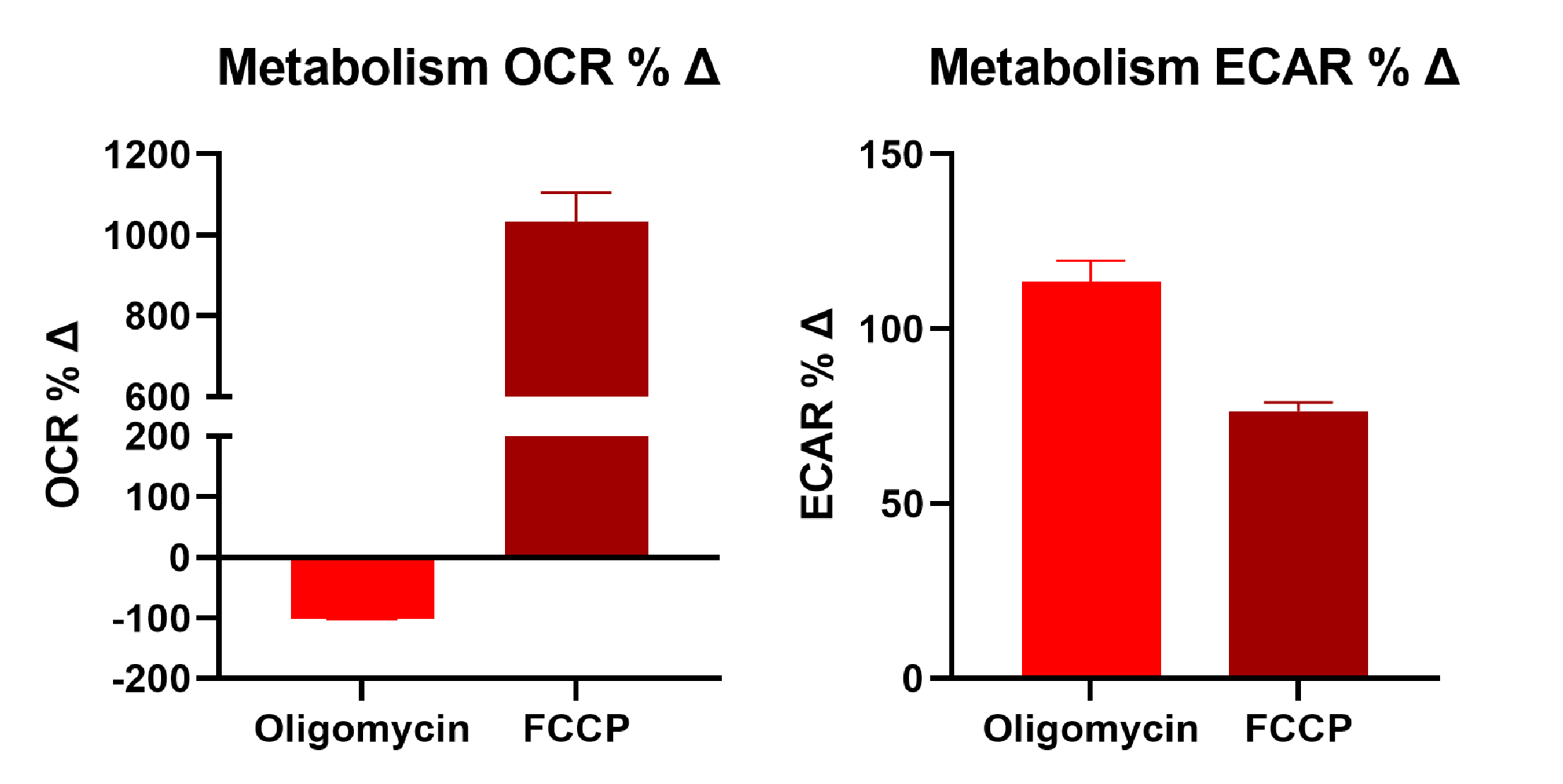 Oligomycin inhibits ATP synthase resulting in reduced mitochondrial respiration (OCR) and increase glycolysis (ECAR). Carbonyl cyanide-4 phenylhydrazone (FCCP) collapses the proton gradient and disrupts mitochondrial membrane potential increasing oxygen consumption (OCR) with no or little effect on glycolysis (ECAR).
Assessing mitochondrial depolarization in Ncyte CNS neurons to quantify cytotoxic damage of compounds.
Mitochondrial membrane potential controls respiratory rate, ATP production and generation of reactive oxygen species, which are essential processes for cell homeostasis. Our scientists use confocal microscopy and JC-1 dye as a sensitive readout of mitochondrial depolarization and disruption after treatment with the therapeutic compounds of interest.
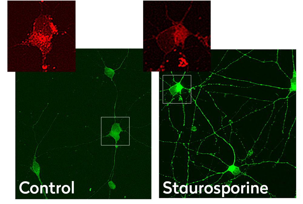 Confocal images of Ncyte CNS neurons stained with JC-1. Emission at 530 nm (green) and 590nm (red) in control and treated cells. Staurosporine treatment reduces mitochondria puncta pattern and depolarization.
 JC-1 590/530 nm emission ratio in Ncyte CNS neurons treated with increasing concentrations of Staurosporine (apoptosis inducer).
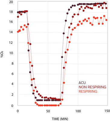
Assessment of oxidative stress levels in Ncyte Cardiomyocytes to measure changes in cardiac metabolism during ischemia/reperfusion.
During myocardial infarction, oxygen supply to cardiac tissue is reduced and it is rapidly restored after reperfusion treatment, inducing changes in cell metabolism. Our scientists can model ischemia/reperfusion on human iPSC-derived cardiomyocytes by using an atmospheric control unit (ACU) and measure oxygen consumption in real time to study cells’ metabolism under different environmental conditions.
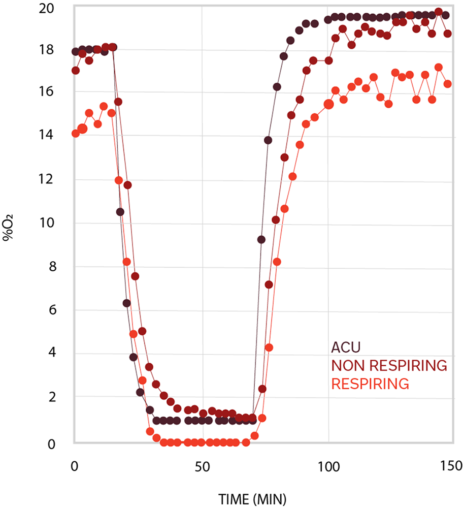 Cell oxygenation traces from cardiomyocytes with oxygen consumption (light red) and from cardiomyocytes in hypoxia (dark red). The black trace represents the oxygen level inside the chamber.
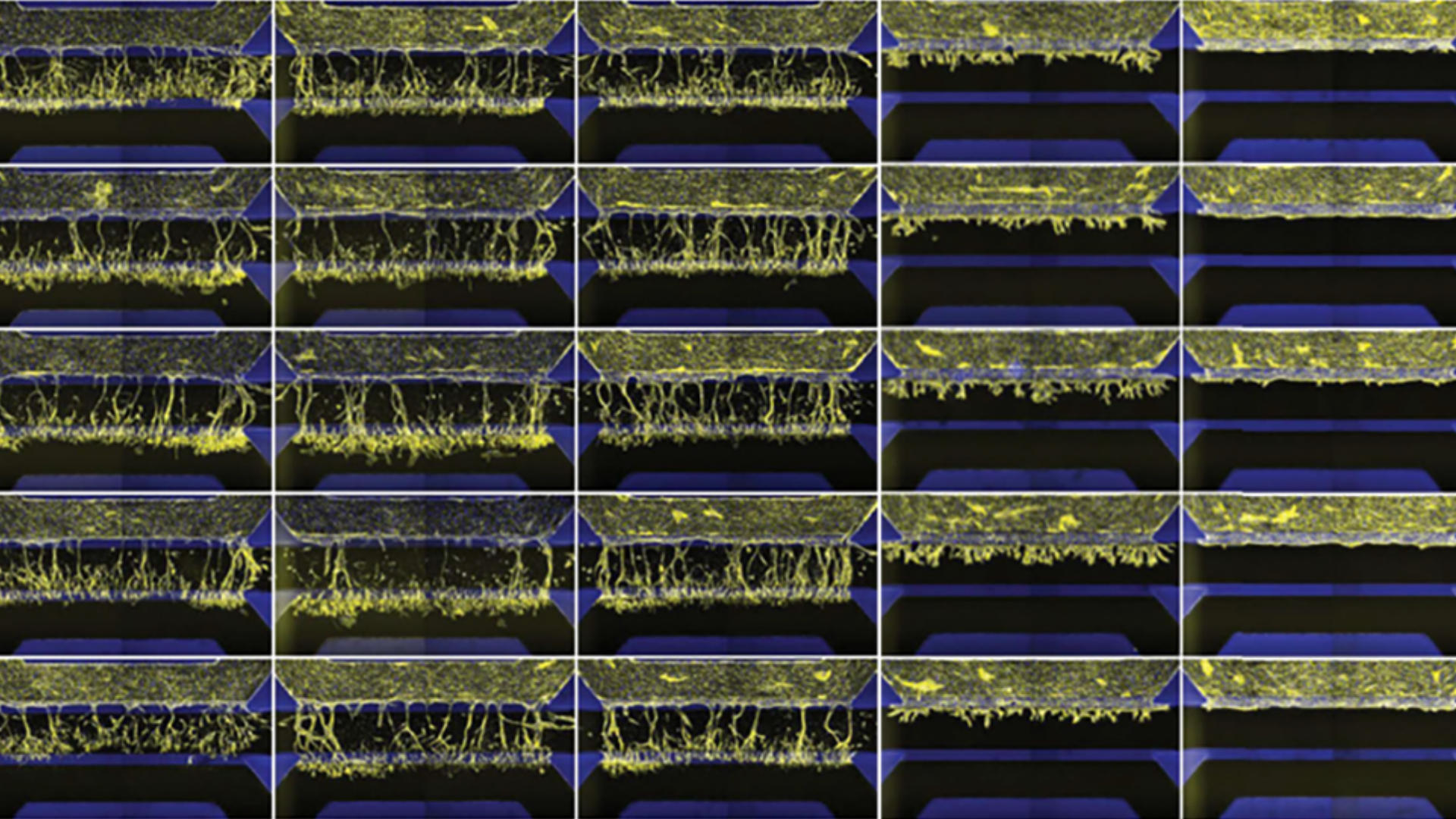
Study compound-induced effects on blood vessel formation
Assay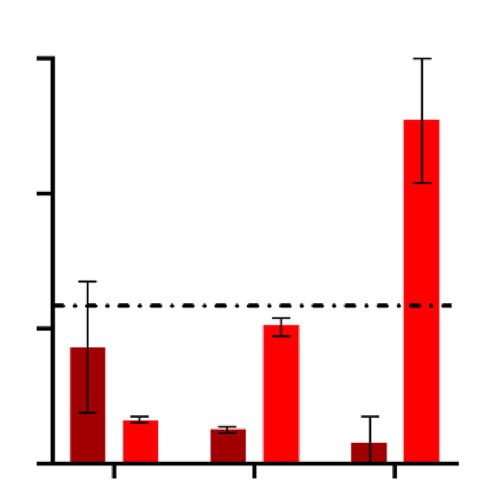
Evaluate and quantify the levels of clinically relevant biomarkers
Assay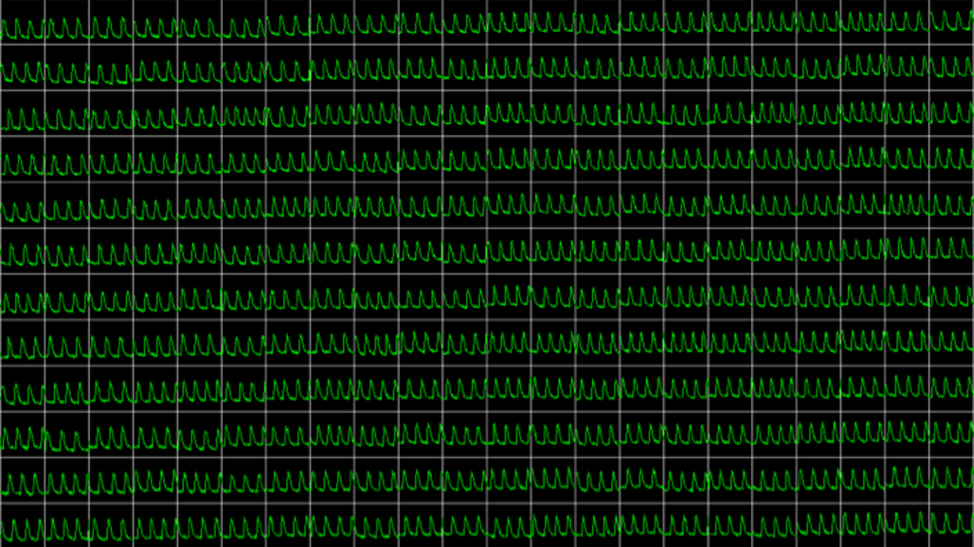
Obtain real-time recordings of intracellular calcium fluctuations
Assay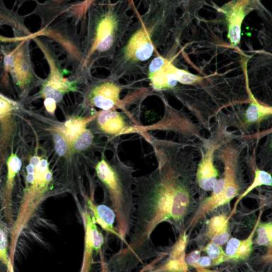
Get in-depth and unbiased insights into the effects of therapeutic candidates on cells
Assay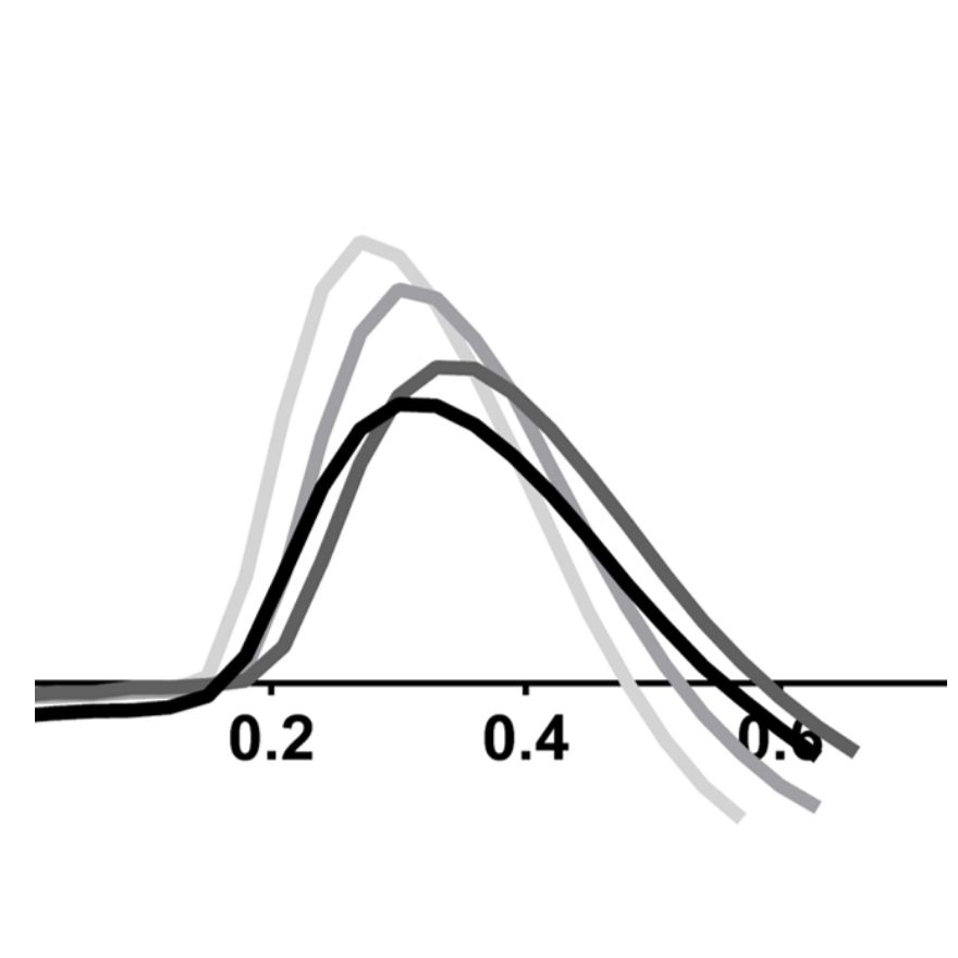
Study drug-induced effects on contractility of cardiac or skeletal muscle cells
Assay
Determine the electrophysiological effects of your therapeutic candidates
Assay
Evaluate endothelial permeability in short and long term
Assay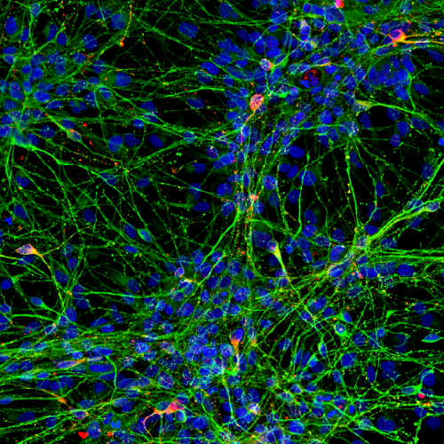
Study drug-induced phenotypic changes in the cell model of your interest
Assay
Assess functional toxicity across multiple human iPSC-derived tissues in a single integrated workflow to support safer IO development decisions.
AssayObtain precise information on compounds' impact on metabolic processes